Table of Contents
What are the Side Effects of Laser Pigmentation Removal in Hyderabad?
Facial hyperpigmentation—whether presenting as stubborn melasma, dark sun spots, or post-acne marks—is an exceptionally common concern among Indian women and men. As energy-based dermatological procedures become increasingly accessible across Telangana, patients frequently inquire about the potential side effects of laser pigmentation removal before undergoing treatment.
While laser technology offers a precise, non-surgical method for shattering excess dermal and epidermal melanin, skin native to the Indian subcontinent possesses distinct biological traits that alter how laser energy is absorbed. Understanding the normal post-procedure healing responses versus adverse clinical side effects allows patients in Hyderabad to make informed decisions and choose practitioners who prioritize skin barrier safety.
What is Laser Pigmentation Removal and What are Its Side Effects?
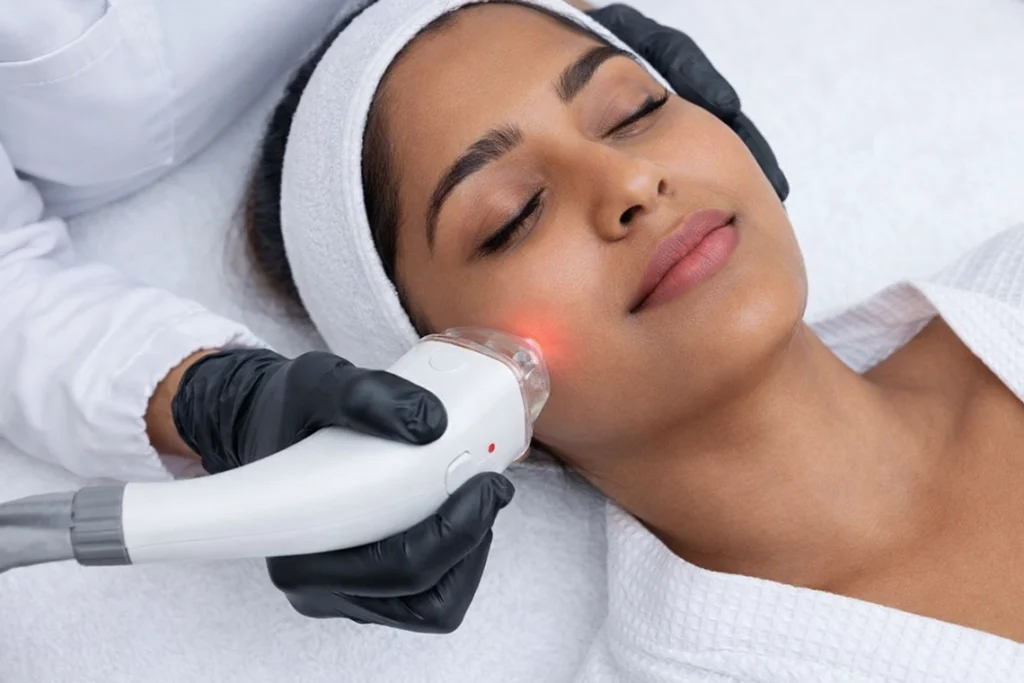
Laser pigmentation removal uses targeted light energy (such as 1064 nm Q-switched Nd:YAG lasers) to fragment excess melanin deposits (ICD-10: L81.8) in the skin. Expected side effects include transient erythema (redness) and mild edema (swelling) lasting 24–48 hours. Adverse risks in darker skin types include post-inflammatory hyperpigmentation (PIH), spotty hypopigmentation, crusting, thermal micro-burns, and rebound melasma flare-ups.
Laser–Skin Interaction and Possible Side Effects
Laser treatments for pigmentation use light energy to target melanin in the skin. The skin’s response depends on the laser type, wavelength, settings, treatment technique, and individual skin characteristics. Skin types III–V may need particular care because inflammation can trigger pigmentation changes.
| Treatment Response | What It Means |
|---|---|
| Mild, Temporary Redness | Short-term redness may occur after treatment. |
| Perifollicular Swelling | Small areas of swelling around hair follicles may develop with certain laser treatments. |
| Surface Micro-Crusting | Fine crusting may occur after some procedures, depending on the treatment and skin response. |
Potential Adverse Effects
| Side Effect | Description |
|---|---|
| Post-Inflammatory Hyperpigmentation (PIH) | Dark marks may develop after treatment-related inflammation or skin injury. |
| Hypopigmentation | Areas of skin may become lighter than the surrounding skin. Small, drop-like patches are sometimes described as guttate hypopigmentation. |
| Melasma Flare or Recurrence | Melasma may worsen or return after treatment, particularly if the skin becomes inflamed or receives excessive UV exposure. |
| Burns or Scarring | Excessive energy or inappropriate treatment can cause tissue injury and, in some cases, scarring. |
Why Does Indian Skin React Differently to Laser Energy?
Evaluating laser safety requires an understanding of the physiological characteristics of Indian skin. Complexions native to the Indian subcontinent typically fall between Fitzpatrick Scale Types III and V. This skin classification features a high concentration of active, melanocytes—the pigment-producing cells located along the basal layer of the epidermis.
Lighter Skin vs. Darker Skin: Melanin, Sunburn, and Pigmentation Risk
Melanin helps protect the skin from some effects of ultraviolet (UV) radiation. However, sunburn and post-inflammatory hyperpigmentation (PIH) can occur in all skin types, and the risk varies between individuals.
| Feature | Lighter Skin (Fitzpatrick Types I–II) | Darker Skin (Fitzpatrick Types III–V) |
|---|---|---|
| Melanin Levels | Generally lower | Generally higher |
| Sunburn Risk | More likely to burn easily | May burn less easily, but sunburn is still possible |
| PIH Risk | Can develop dark marks after acne, irritation, or injury | Dark marks may be more noticeable and persist longer |
| Laser Treatment Considerations | Treatment still requires appropriate settings and sun protection | Careful device selection and individualised settings are important to reduce pigmentation complications |
In Fitzpatrick Types III–V skin, epidermal melanin acts as a competing chromophore. When a laser fires, epidermal melanin competes with deeper target pigment spots for light energy absorption. If laser energy (fluence) or thermal heat is delivered too aggressively, epidermal cells experience thermal injury. This releases inflammatory mediators that trigger surrounding melanocytes to produce excess melanin, resulting in post-inflammatory hyperpigmentation (PIH).
Regional environmental and lifestyle factors in Hyderabad compound these physiological risks:
- High Solar UV Exposure: Hyderabad experiences high UV index levels throughout the year. Exposing laser-treated skin to ambient solar radiation without adequate photoprotection significantly increases the risk of post-procedure hyperpigmentation.
- Humid Summer and Monsoon Heat: Hot, humid environmental conditions induce facial vasodilation (dilation of blood vessels). Thermal heat from the environment combined with residual laser heat increases tissue inflammation.
- Occupational Blue Light and Digital Screen Exposure: Tech professionals in Madhapur, Gachibowli, and HITEC City face prolonged High-Energy Visible (HEV) blue light exposure. Blue light stimulates opsin-3 receptors on melanocytes, aggravating post-laser pigmentation if skin is left unshielded.
What Causes Adverse Side Effects During Laser Pigmentation Treatment?
Adverse outcomes from laser treatments rarely occur at random; they stem from specific physical, clinical, and procedural variables:
- High Fluence or Inappropriate Wavelength Selection: Utilizing short wavelengths (such as 532 nm) at high energy settings on Fitzpatrick Types IV–V skin causes excessive superficial thermal damage rather than fragmenting deeper dermal pigment safely.
- Thermal Heat Accumulation: Using continuous-wave or long-pulse lasers instead of photo-acoustic Q-switched or picosecond platforms causes thermal energy to spill into surrounding tissue, causing collateral thermal burns.
- Lack of Pre-Treatment Skin Priming: Initiating laser sessions on active melanocytes without 2 to 4 weeks of topical priming (using tyrosinase inhibitors like azelaic acid or kojic acid) leaves pigment cells unstable and prone to reactive hyperpigmentation.
- Inadequate Post-Procedure Photoprotection: Skipping broad-spectrum sunscreen or exposing newly treated skin to direct sunlight triggers rapid melanin synthesis in vulnerable basal cells.
- Aggressive Friction and Mechanical Exfoliation: Scrubbing or picking at micro-crusts following a laser session tears fragile epidermal tissue, creating micro-wounds that heal as dark spots.
- Uncontrolled Hormonal Triggers: Treating vascular melasma without addressing underlying hormonal shifts (such as PCOS, oral contraceptive use, or thyroid dysfunction) can trigger severe rebound melasma flares due to laser-induced vascular stimulation.
How Do Normal Reactions vs. Adverse Side Effects Look and Feel?
Distinguishing between normal post-laser healing and clinical complications is essential for patient safety:
- Transient Erythema and Perifollicular Edema (Normal): Mild pinkness or redness accompanied by slight swelling around hair follicles that subsides naturally within 2 to 24 hours post-session.
- Mild Frosting or Surface Micro-Crusting (Normal): A faint, temporary whitening of targeted spots immediately following laser impact, followed by tiny, sand-like micro-crusts that flake off naturally over 3 to 7 days.
- Post-Inflammatory Hyperpigmentation (Adverse): Broad, dark brown patches developing across the treated area 1 to 3 weeks after the procedure, making the treated skin darker than it was before the laser session.
- Confetti-like or Guttate Hypopigmentation (Adverse): Small, permanent white spots where melanocytes have been thermally destroyed by excessive energy, leaving patches devoid of natural skin pigment.
- Blistering, Pinpoint Bleeding, or Scarring (Adverse): Surface fluid-filled vesicles or crusting caused by excessive thermal energy, indicating thermal damage to the dermal-epidermal junction.
What Can Make Laser Pigmentation Side Effects Worse in Indian Skin?
Unintentional home care habits and improper post-procedure choices can significantly increase complication rates:
Post-Laser Factors That May Delay Skin Recovery
| Avoid | Why It Matters |
|---|---|
| ☐ Picking or scratching micro-crusts | May delay healing and increase the risk of scarring or post-inflammatory hyperpigmentation (PIH). |
| ☐ Hot showers, saunas, and excessive heat | Can worsen temporary redness, flushing, and irritation after laser treatment. |
| ☐ Using strong acid serums too soon | Ingredients such as glycolic acid, salicylic acid, and retinoids may irritate the healing skin barrier. |
| ☐ Skipping sun protection | UV exposure can worsen pigmentation. Tinted sunscreen containing iron oxides may also help protect against visible light that can contribute to pigmentation, particularly in darker skin tones. |
| ☐ Applying unsterilized traditional oils or home remedies | These products may irritate treated skin or introduce contaminants; some may also be unsuitable for the healing area. |
- Picking or Scratching Flaking Micro-Crusts: Manually peeling tiny crusts off targeted spots tears immature epidermal tissue underneath, causing permanent textural scarring and hyperpigmentation.
- Exposing Treated Skin to Direct Thermal Heat: Taking steam baths, using saunas, cooking over open heat, or washing with hot water within 48 hours prolongs cutaneous vasodilation and inflammatory cytokine release.
- Applying Exfoliating Active Serums Too Soon: Using high-concentration glycolic acid, salicylic acid, or topical retinoids immediately after a laser session disrupts the healing skin barrier, causing severe chemical contact dermatitis.
- Using Non-Tinted Sunscreens: Applying clear sunscreens that lack iron oxides fails to reflect HEV blue light and visible light, leaving pigment cells vulnerable to indoor and outdoor darkening.
- Applying Thick, Unsterilized Home Oils: Coating warm, laser-treated skin with heavy coconut or mustard oil traps thermal heat within the dermis, escalating tissue inflammation.
Step-by-Step Clinical Protocol: Minimizing Laser Side Effects
Board-certified dermatologists follow a strict 5-step clinical protocol to ensure safety and minimize complications when performing laser pigmentation treatments on Indian skin:
Step-by-Step Approach to Pigmentation Treatment
| Stage | Approach | Purpose |
|---|---|---|
| 1. Diagnosis | Dermoscopy, Wood’s lamp examination when appropriate, and assessment of pigment depth | Helps evaluate the type and distribution of pigmentation. These tools have limitations and may not reliably determine pigment depth in every case. |
| 2. Skin Priming | Consider topical tyrosinase inhibitors for an appropriate period, often 2–4 weeks if prescribed | May help regulate melanin production and prepare the skin for selected procedures. The product and duration depend on the diagnosis and skin sensitivity. |
| 3. Patch Testing | Test a conservative treatment setting on a small area and assess the response | Helps the clinician evaluate tolerability and identify potential adverse reactions before broader treatment. A 48-hour check alone may not detect every delayed reaction. |
| 4. Low-Fluence Laser Care | Consider low-fluence Q-switched laser treatment when clinically appropriate, with suitable cooling and protective measures | Aims to target selected pigment while limiting unnecessary irritation. Laser treatment can also worsen pigmentation, particularly in skin prone to post-inflammatory hyperpigmentation (PIH). |
| 5. Maintenance | Use suitable non-hydroquinone topical treatments when indicated, plus broad-spectrum tinted sunscreen with iron oxides, SPF 30–50 | Helps maintain results and reduce UV- and visible-light-related pigmentation. Reapply sunscreen as directed, especially during prolonged outdoor exposure. |
- Step 1: Diagnostic Classification and Pigment Mapping: Utilizing dermoscopy and Wood’s lamp examination to determine pigment depth (epidermal, dermal, or mixed) and evaluate baseline vascular inflammation.
- Step 2: Pre-Procedure Melanocyte Priming (2 to 4 Weeks): Applying non-hydroquinone tyrosinase inhibitors (such as azelaic acid, kojic acid, or arbutin) to stabilize pigment-producing cells prior to laser exposure.
- Step 3: Spot Patch Testing: Applying conservative test spots along the jawline 48 to 72 hours before full-face treatment to evaluate individual tissue response and check for hyper-reactivity.
- Step 4: Low-Fluence Q-Switched Nd:YAG Energy Application: Utilizing 1064 nm wavelength energy in nanosecond or picosecond pulse durations with continuous forced-air cooling to fragment pigment acoustically rather than thermally. [INTERNAL LINK: laser toning for pigmentation]
- Step 5: Post-Procedure Barrier Repair and Photoprotection: Applying soothing, barrier-repairing formulations containing ceramides or centella asiatica alongside broad-spectrum tinted mineral sunscreens containing iron oxides.
Comparison: Laser Technologies and Their Safety Profiles in Indian Skin
The table below compares common laser platforms used for pigmentation removal, their primary mechanisms, safety profiles for Fitzpatrick Types III–V skin, and risk levels for post-inflammatory hyperpigmentation:
| Laser Platform & Wavelength | Primary Mechanism | Primary Indications | PIH Risk Level (Indian Skin) | Safety & Recovery Profile |
| Q-Switched Nd:YAG (1064 nm) | Photo-acoustic energy fragments dermal and epidermal melanin without excessive heat. | Dermal melasma, post-acne marks (PIH), freckles, sun spots. [INTERNAL LINK: GFC hair therapy] | Low (When used with low-fluence laser toning technique). | Minimal downtime; mild transient redness lasting 1–2 hours. |
| Picosecond Laser (1064 nm) | Ultra-short picosecond pulses break pigment into tiny dust-like particles using pressure waves. | Recalcitrant melasma, stubborn tattoos, deep dermal spots. | Very Low (Photomechanical impact minimizes heat transfer). | Rapid recovery; mild erythema subsides within hours. |
| Fractional Er:YAG Laser (2940 nm) | Creates microscopic ablative channels to resurface epidermal tissue. | Textural roughness, epidermal spots, superficial acne scars. | Moderate to High (Requires pre-priming and barrier care). | 3–5 days of micro-crusting, bronzing, and light peeling. |
| Intense Pulsed Light (IPL / 500–1200 nm) | Broad-spectrum light energy targets melanin and vascular structures simultaneously. | Superficial freckles, sun damage in fair complexions. | High in Darker Tones (Broad light spectrum risks thermal burns). | Requires cautious parameter selection in Fitzpatrick IV–V skin. |
| Q-Switched Frequency-Doubled (532 nm) | High-absorption wavelength targets superficial epidermal melanin aggressively. | Epidermal freckles, lentigines in lighter complexions. | Very High (High epidermal absorption risks thermal injury). | Avoided or used at low settings in darker complexions. |
When to See a Dermatologist in Hyderabad
While mild redness immediately following a laser session is expected, specific warning signs require prompt medical evaluation by a qualified specialist.
Clinical Evaluation: Warning Signs After Laser Treatment
| Warning Sign | What to Look For | Recommended Action |
|---|---|---|
| Darkening of the Skin | Treated areas becoming noticeably darker over the following 1–2 weeks | Contact your treating dermatologist to assess possible post-inflammatory hyperpigmentation (PIH). |
| White Spots | New, sharply defined or confetti-like white patches (possible hypopigmentation) | Arrange a dermatology review to evaluate pigment loss. |
| Blistering or Painful Crusts | Fluid-filled blisters, painful scabs, or skin breakdown | Seek prompt medical assessment; do not pick, peel, or pop the affected areas. |
| Prolonged Redness or Swelling | Redness or swelling that persists beyond 72 hours, worsens, or fails to improve | Contact your treating clinician. The expected recovery period varies by laser type and treatment intensity. |
| Severe or Persistent Burning | Burning discomfort that continues despite gentle cooling or becomes more intense | Stop applying active skincare products and seek prompt medical advice. Avoid placing ice directly on the skin. |
Clinical Practice Highlight:
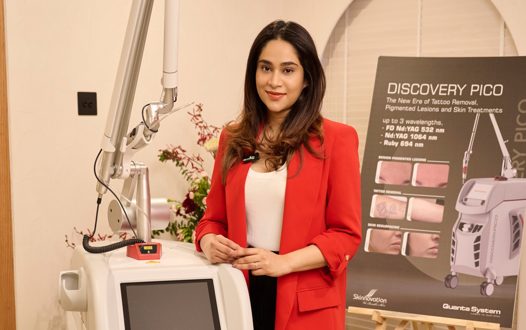
Patients seeking evidence-based laser pigmentation removal and complication management in West Hyderabad can visitDr. Showrya Gadde’s clinic in Kondapur. As a board-certified dermatologist (MBBS, MD Dermatology) with fellowship training in aesthetic medicine, Dr. Gadde utilizes US-FDA approved 1064 nm Q-switched Nd:YAG laser technology and picosecond platforms. Every patient undergoes pre-laser melanocyte priming, dermoscopic evaluation, and low-fluence customized protocols to protect Indian skin from post-laser hyperpigmentation.
Consultations can be scheduled via drshowryagadde.com or WhatsApp +91 7599741111.
Questions to Ask at Your Laser Consultation
Ensure safety and clarity by discussing these key questions with your provider prior to starting treatment:
- “Is the laser platform US-FDA approved and specifically calibrated for Fitzpatrick Type IV–V skin?”Why it matters: Confirms the equipment has undergone safety testing for darker complexions to minimize thermal damage.
- “Will my skin require a pre-treatment priming routine with topical tyrosinase inhibitors?”Why it matters: Pre-priming for 2 to 4 weeks stabilizes melanocyte activity and significantly reduces the risk of post-laser hyperpigmentation.
- “Are you using a photo-acoustic low-fluence laser toning technique or high-heat photothermal energy?”Why it matters: Low-fluence photo-acoustic energy fragments pigment safely without generating excessive heat that could trigger rebound melasma.
- “Will a spot patch test be performed on my jawline prior to full-face treatment?”Why it matters: Allows your provider to observe your skin’s response and adjust energy settings safely.
- “What specific barrier-repair products and tinted sunscreens should I use after my session?”Why it matters: Proper post-procedure care protects healing skin from environmental heat, UV rays, and digital blue light.
Frequently Asked Questions (FAQs)
1. Can laser pigmentation removal make my dark spots permanently worse?
If performed with incorrect laser wavelengths, excessive energy, or without pre-treatment skin priming, lasers can trigger severe post-inflammatory hyperpigmentation (PIH) or rebound melasma in Indian skin. However, when administered by a trained dermatologist using low-fluence Q-switched techniques, the risk of worsening pigmentation is minimized.
2. How long do normal side effects like redness last after a laser toning session?
Normal post-laser reactions—such as mild pinkness (erythema) or slight swelling around hair follicles—typically resolve on their own within 1 to 2 hours following low-fluence Q-switched Nd:YAG laser toning.
3. What is the difference between hyperpigmentation and hypopigmentation post-laser?
Post-laser hyperpigmentation is a temporary darkening of treated skin caused by excess melanin production following heat or inflammation. Hypopigmentation refers to light or white spots caused by thermal destruction of pigment cells (melanocytes), which is much more difficult to treat.
4. Why is broad-spectrum tinted sunscreen mandatory after laser sessions in Hyderabad?
Treated skin remains sensitive to light for several weeks after a laser session. Tinted mineral sunscreens containing iron oxides block ultraviolet rays (UVA/UVB) as well as High-Energy Visible (HEV) blue light, preventing melanocytes from producing reactive melanin.
5. Can I wear makeup or apply active skincare serums immediately after a laser session?
No. You should avoid heavy makeup, physical scrubs, and active exfoliating serums (such as glycolic acid, salicylic acid, or retinol) for 3 to 5 days after a laser treatment to allow the skin barrier to heal without irritation.
6. Are picosecond lasers safer for dark Indian skin tones than traditional nanosecond lasers?
Picosecond lasers deliver energy in ultra-short picosecond pulses, breaking pigment into tiny particles primarily through mechanical pressure waves rather than thermal heat. This reduced thermal impact makes picosecond lasers safer for melanin-rich skin when used correctly.
7. How many laser sessions are usually required to clear dermal melasma safely?
Clearing dermal melasma safely in Indian skin requires a patient, conservative approach. Dermatologists typically recommend 6 to 8 low-fluence laser toning sessions spaced 2 to 4 weeks apart, combined with daily topical maintenance.
8. How do I schedule a pre-laser diagnostic skin evaluation in West Hyderabad?
You can book an assessment with a board-certified dermatologist through official clinic contact channels. For instance, consultations at a specialized practice in West Hyderabad can be requested at drshowryagadde.com or via WhatsApp at +91 7599741111.
Final Thoughts: Prioritizing Safety and Clinical Precision
Understanding the side effects of laser pigmentation removal highlights the importance of choosing qualified medical care. While laser technology provides an effective method for treating hyperpigmentation, melanin-rich Indian skin requires conservative settings, careful pre-treatment priming, and diligent post-procedure care.
Prioritize treatments led by board-certified dermatologists who utilize US-FDA cleared Q-switched or picosecond platforms, perform pre-laser patch testing, and emphasize skin barrier health. Taking an evidence-based approach ensures your skin receives safe, effective treatment for long-term clarity.

Dr. Showrya Gadde, MBBS, MD (Dermatology), is a board-certified dermatologist and fellowship-trained aesthetic clinician in Kondapur, Hyderabad. She is best known for pico laser, Cosmelan peel, Botox and acne scar treatments. Every treatment she offers is evidence-based and customised for Indian skin.